8 April 2024
Cat mummy CT image rendered with Dragonfly software (v.2022, Comet Technologies Canada Inc.)
Summary
What do skeletal biologists, biological anthropologists, mechanical engineers and museum curators have in common? Perhaps, surprisingly, a fascination with ancient Egyptian mummies! In 2023, researchers Natalie Reznikov from McGill University, and Andrew Nelson from Western University, approached McGill's Redpath Museum curators Annie Lussier and Anthony Howell with a request to analyze animal mummies from their World Cultures collection. Seven fragile wrapped bundles—presumably containing two hawks, two crocodiles, one cat and two ibises—were carefully delivered to the new X-ray imaging facility at ÉTS, to be investigated in collaboration with Professor Vladimir Brailovski.
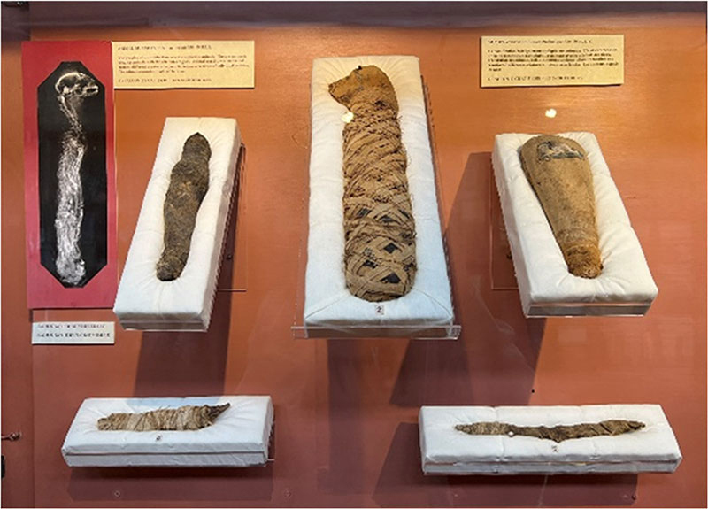
Figure 1. Mummified animals displayed in the Ancient Egypt section of the Redpath Museum.
Mummified Remains
For thousands of years, mummifying dead bodies was one of the main religious practices of the ancient Egyptian civilization. Artificial mummification involved embalming the body with resin, desiccation, and careful wrapping, to preserve it “intact” for the afterlife. Royals and aristocrats, clerics and sacred animals, and even pets and food offerings were all mummified. Mummification relied on substantial knowledge of chemistry and anatomy, and was perfected over many years through a blending of science and art. But it was also a trade: animals of cultural significance were commonly preserved as symbolic offerings for deities. Such preparations of birds, mammals and reptiles are now called “votive” mummies (Figures 1 and 2). These were among the most numerous mummies in the late Ptolemaic period: a citizen could purchase a mummy and bring it to a temple, hoping to secure a personal favour from the relevant god, just like a contemporary believer would light a candle in a church. A different deity and a different favour to ask would require a different votive mummy.
Animal Mummies in the Redpath Museum
The seven animal mummies at the Redpath Museum are all votive mummies, dating from more than two thousand years. They are all lightweight and delicate, with a subtle earthy-brown colour, and must be handled with the utmost care. The cat bundle is the largest: the criss-cross ribbon wrapping is regular and ornate, and characteristic pointy ears are made of folded textile (Figure 2a). Both juvenile crocodile bundles are long and narrow, one with its distinctive 'grinning' jaws sticking out of the rough cloth (Figure 2b), and the other with gracile bony knees splaying out from the textile layers (Figure 2c) as if they had been hastily wound. The hawks are solemn and compact, both with their beaks peaking out through the wrappings (Figures 2d and 2e). The two ibis bundles are about 12 inches (30 cm) long, and have a characteristic pear shape, with a bulging head end. This wading long-legged bird was highly revered in ancient Egypt as a symbol of literacy and wisdom, and also as protection for farmers against snakes and plague. One of the pear-shaped bundles has a plaque with a picture of an ibis painted on it with white, black and blue mineral dyes (Figure 2f). The other is tightly wrapped in a criss-cross pattern of ribbons (Figure 2g).
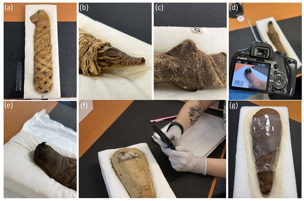
Figure 2. Votive mummies: a) Cat 2728.01, b) Crocodile 2725.01, c) Crocodile 2725.02, d) Falcon 2726.02, e) Falcon 5731, f) Ibis 2727.01, and g) Ibis 2727.02.
Transportation and Mounting
The bundles must be transported by a professional museologist (Josée Noël, CARTGO) in custom-made cushioned cradles (Figure 3). Doctoral student Shumeng Jia designed and 3D-printed an inclined support for the cradles. This sturdy fixture is now used to mount and stabilize the precious bundles on a rotating platform inside the X-ray tomographic scanner. Shumeng and MA student of anthropology Maris Schneider, both wearing gloves and masks to protect the specimens, carefully centre the inclined adaptor with the mummy in the padded cradle inside the futuristically lit chamber of the X-ray tomography scanner. It is now time to perform the scan. Salah Brika, a postdoctoral fellow, shuts the sliding door of the scanner and turns on the X-rays. Everybody crowds around the control station as the first hazy, see-through image appears on the screen.
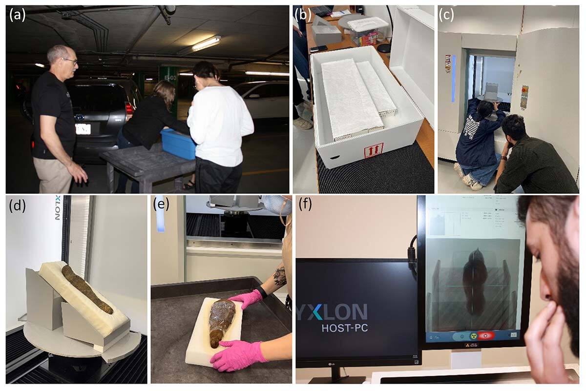
Figure 3. Handling the mummies. a) Mummy transport, b) Mummy package, c) Placing the mummy holder, d) Falcon mummy in holder on the rotating platform, e) Preparing the ibis mummy, f) Image appears.
X-Ray Micro-Computed Tomography
The imaging technique known as X-ray micro-computed tomography (micro-CT) uses X-ray technology to create detailed cross-sectional and volumetric images of an object. It is widely used in medicine to provide information about the internal structure of organs, especially bones and teeth. It is also used in industrial applications for nondestructive testing and quality control. The micro-CT scanner consists of an X-ray source and a detector array mounted opposite each other (Figure 4). The X-ray source emits a cone-shaped beam, and the detector array captures the attenuated X-rays passing through the object. In micro-CT, the object is rotated while the X-ray source and the detector are stationary (in a medical CT scanner, the X-ray source and detector rotate around the patient during imaging). Every time the platform turns by one-tenth of a degree, an X-ray projection image is taken. Since X-rays can pass through textiles and resin, but scatter when they encounter bone or sediment, the inner structure of the bundles can be revealed through contrast differences. The raw X-ray projections collected from multiple angles are processed by a computer to render a 3D digital image—this is known as volume reconstruction. The reconstructed images are composed of voxels, which are three-dimensional pixels. Each voxel represents a small volume element in the object and has a corresponding density value based on the degree of X-ray attenuation.
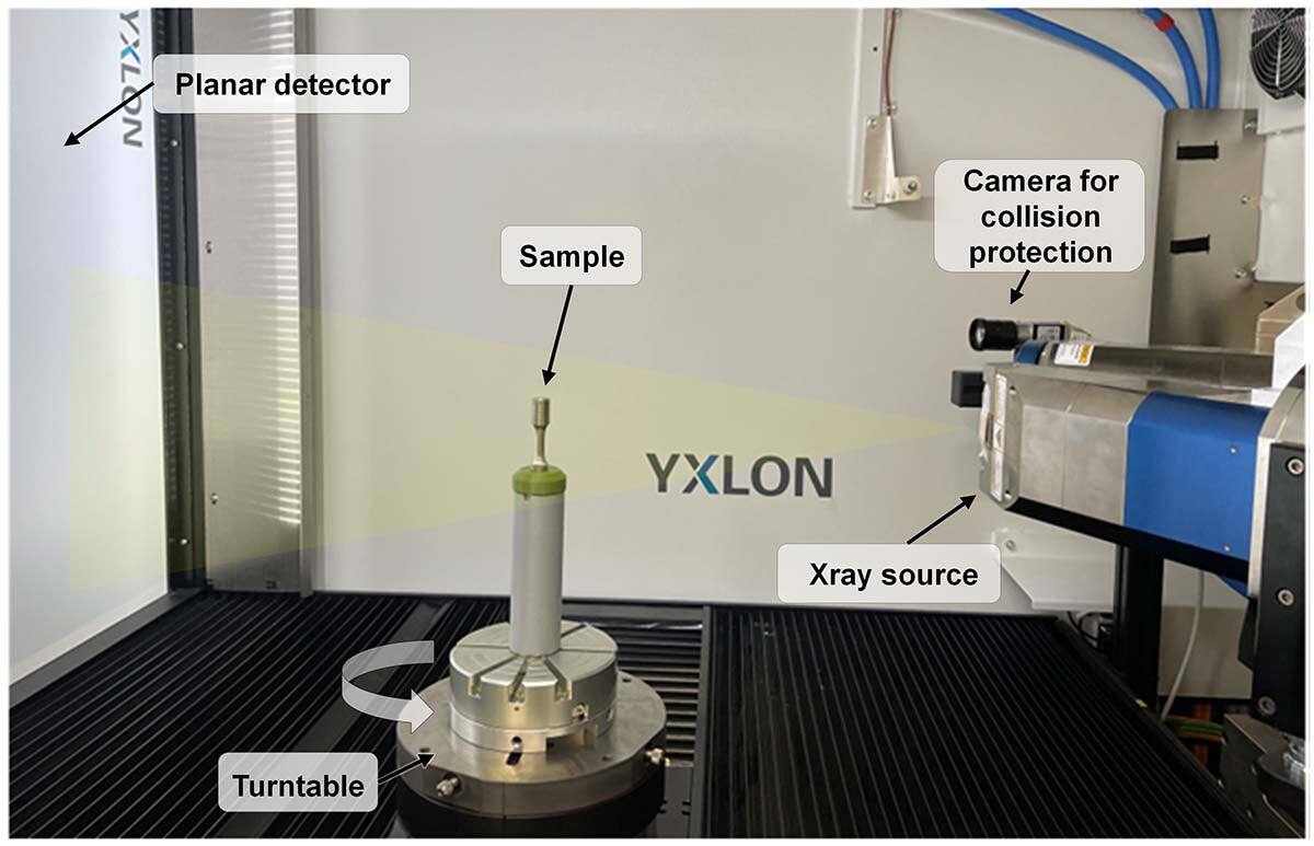
Figure 4. Configuration of the X-ray micro-CT scanner (Yxlon FF35).
3D Digital Reconstruction
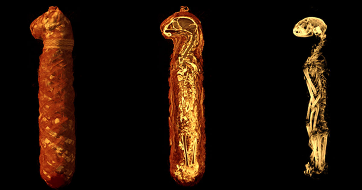
The images are steadily collected and provisionally reconstructed as they appear on the screen. While most of the image analysis work will be done later, some features are already obvious. The cat’s bones under the elegant bandages are severely dislocated, and its small brain has dried to dust inside the skull (Figure 5).
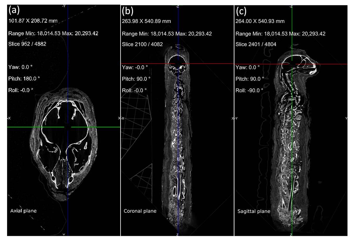
Figure 5. Micro-CT scans of cat mummy 2728.01 in a) axial, b) coronal, and c) sagittal views.
The baby crocodiles are tiny and endearing—yet it remains to be determined whether they are juveniles of the fierce Nile crocodile, or of another species. In one of the crocodiles, the team notices an additional tiny bone near the tail that is not normally part of a crocodile (Figure 6). What is it? It appears to have been accidentally lodged into this bundle by a busy, inattentive embalmer on the 'votive mummy production line.' The hawks look very similar to one another—the quills of their primary feathers are well preserved, and both of their windpipes (trachea) are intact (Figure 7).
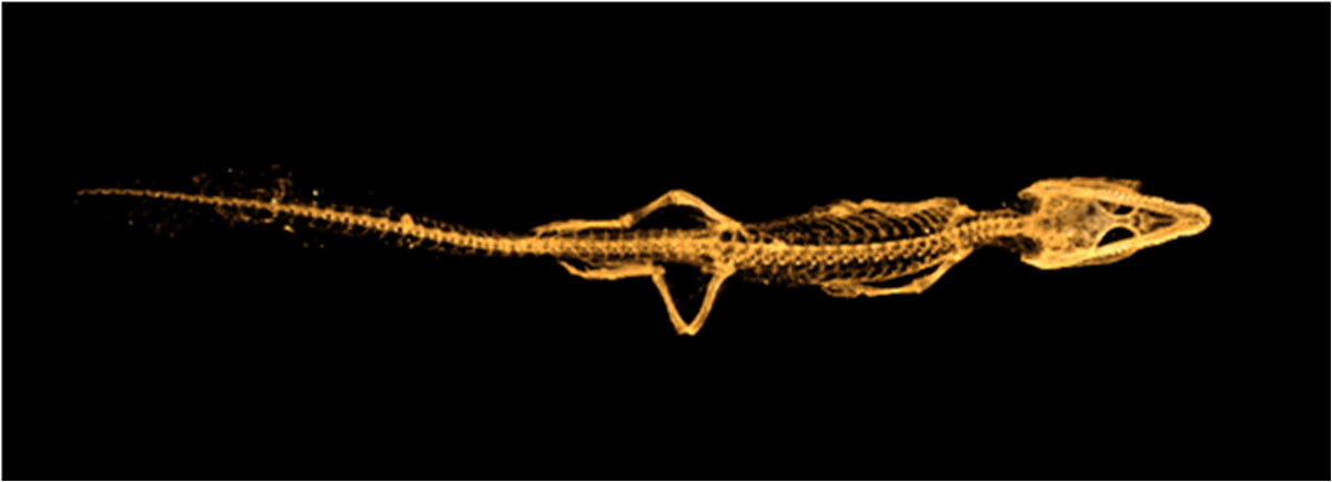
Figure 6. Skeleton of juvenile crocodile mummy 2725.02.
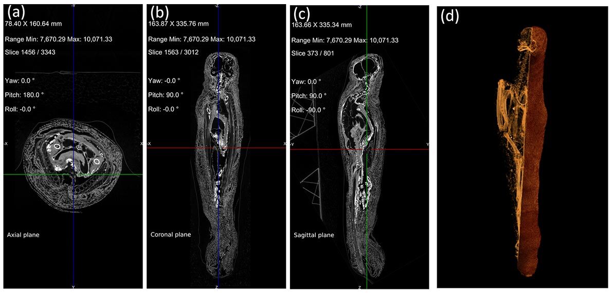
Figure 7. Micro-CT scan of falcon mummy 5731 in a) axial, b) coronal and c) sagittal views, and d) 3D rendering of falcon mummy 2726.02.
A Pseudo-Mummy
Next, when the first X-ray projection image of one ibis bundle emerges on the screen, the team is perplexed. They try to adjust the brightness and contrast to see more clearly, but nothing helps to show the skeleton. Alas, and quite surprisingly, inside the first ibis 'mummy' there is in fact no mummy! There are only packed rags partially smeared with clay and dirt, shaped as a votive mummy. There are no bones, and the only things scattering the X-ray beam are small pebbles (Figure 8). This is a pseudo-mummy. But this doesn’t mean that the bundle is not ancient and important. It turns out that it was not unusual for ancient Egyptians to make pseudo-mummies containing only a few random objects or nothing at all. Perhaps, more than two thousand years ago, an unknown artisan sold a fellow citizen an empty bundle shaped as a sacred ibis, to be deposited in the temple.
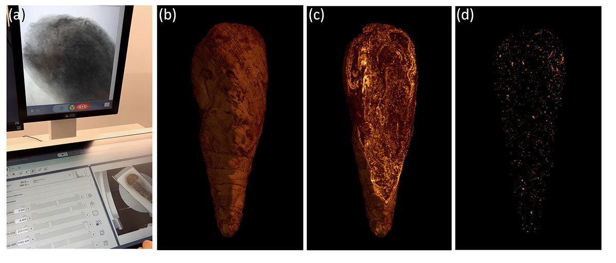
Figure 8. A pseudo-mummy. a) X-ray projection images of ibis mummy 2727.02, b) textile, c) digital section, and d) pebbles.
Will the team have more luck with the second ibis mummy? The painted plaque attached to the exterior of the bundle suggests so, as it features a recognizable image of a gracefully sitting, adult bird with its downward-curved beak and long legs. As the suspense grows, the second bundle does indeed show some bones, although not exactly what the team expected. It’s a hatchling chick, less than the size of a table egg. The characteristic shape of the beak clearly indicates that it is indeed an ibis, however, not the majestic embodiment of Thoth, the god of literacy, but a small baby (Figure 9).
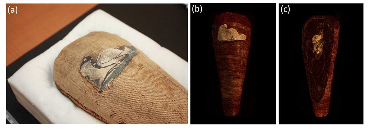
Figure 9. Ibis mummy. a) Painted plaque on ibis mummy 2727.01, b) 3D bundle rendering, c) mummy 2727.01 containing a hatchling ibis.
More to Follow!
Further work is necessary to determine the exact species and cause of death to better understand the cultural context of these preparations. Thanks to the thousands of images acquired over multiple magnifications, the researchers will now be busy for several more years digitally 'unwrapping' the mummies, analyzing them bone-by-bone, and comparing them with modern animals. But the revelation of one mummy being a counterfeit, and the other one being a near-counterfeit, illustrates from this ancient civilization fundamental human characteristics and the alluring call to ‘cut corners.’ Maybe it is fortunate, as at least one ibis got spared. Or maybe not, as Nelson jokes: perhaps, "Many of humanity's misfortunes are the result of the wrath of Thoth, brought about in response to a fake offering?”
____
This study highlights the remarkable versatility of the X-ray micro-computed tomography technique, which can be used to transcend diverse fields. It enables i) non-destructive inspection of metallic parts for detection of defects and for dimensional analysis, ii) thorough examination of intricate electronic assemblies for fault diagnosis and reliability assessment, iii) unveiling of subsurface feature distribution and structural intricacies of concrete and soil samples, and iv) visualizing complex internal structures of biological specimens. Finally, it unravels the untold stories of ancient artifacts.
The versatility of the X-ray micro-computed tomography technique as used across these disciplines underscores its indispensability as a tool for innovation and advancement in various fields. To fully harness its potential, ongoing efforts to refine data by processing algorithms are essential to ensure maximal utilization of its capabilities in different application areas.